
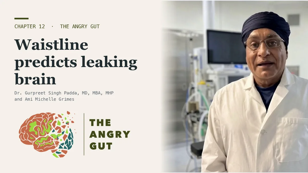
For decades the blood-brain barrier was drawn as a brick wall with the brain safe behind it. I drew it that way for residents. Then researchers put living people in a scanner, tracked contrast dye seeping out of brain blood vessels, and found the wall leaking in one place first.
That place is the hippocampus, where memories are filed, and what opens it looks more like metabolic disease than amyloid. That is the real story behind a leaky brain. The video above comes from Chapter 12 of The Angry Gut, co-written by Ami Michelle Grimes and Dr. Gurpreet Singh Padda, MD, MBA, MHP. This page is about measurement and the numbers you can move.
The barrier leaks first where memories are filed
Using dynamic contrast MRI, one team compared older adults who still had normal cognition against young controls. Permeability was higher by 41% in the hippocampus, 107% in its CA1 subfield and 48% in the dentate gyrus. In mild cognitive impairment, those same regions climbed again against people of the same age, by 24%, 53% and 27% (Montagne et al., 2015).
How do we know the scanner is not just picking up noise? The same series checked people with multiple sclerosis, and their map was the reverse: permeability rose in total white matter, corpus callosum and internal capsule by 32%, 26% and 23%, with no change in the hippocampus. A method that draws a different map in a different disease is measuring something regional and real.
Genetics sharpens the picture. In 245 people scanned and 350 with spinal fluid measured, APOE4 carriers who remained cognitively normal already showed breakdown in the hippocampus and the neighboring parahippocampal gyrus, while amyloid and tau tracer uptake did not differ. Injury to pericytes, the cells that keep the barrier’s seams closed, predicted later decline in carriers and in no one else (Montagne et al., 2020).
The leak shows up before the inflammation
Here is the result almost nobody prints. In the people whose hippocampus was leaking, spinal fluid was checked for the usual inflammatory suspects. Interleukin-6, tumor necrosis factor-alpha, interferon-gamma, adhesion molecules, tau and amyloid: none had moved. Two things did. A marker of pericyte injury was 115% higher in mild impairment than in age-matched controls, and the older albumin ratio was up 30% (Montagne et al., 2015).
A follow-up in 161 people with spinal fluid found no differences between impaired and unimpaired people across 20 inflammatory and degeneration biomarkers, while the barrier measure still predicted impairment after accounting for amyloid and tau (Nation et al., 2019). That cohort excluded vascular dementia, and these papers come from one group with overlapping cohorts: a deepening record, not independent replication.
The order still matters. If the door opens before the inflammation arrives, an anti-inflammatory is late by design. The earlier lever is whatever is prying the door open.
What opens a leaky brain: insulin, blood vessels and body shape
The cheaper, older test is the ratio of albumin in spinal fluid to albumin in blood. In 1,015 people it was unchanged in preclinical and prodromal Alzheimer’s disease and did not follow amyloid imaging or APOE genotype. It followed diabetes, and it rose alongside proteins a strained blood-vessel lining sheds, including vascular endothelial growth factor and two adhesion molecules (Janelidze et al., 2017). In healthy older people from that series, high body mass index and waist-hip ratio predicted a leaking barrier twenty years later.
A second cohort of 1,861 people with dementia found the ratio raised across Lewy body, late-onset Alzheimer’s, vascular and mixed dementia. Within those groups it tracked neurofilament light, a marker of damaged nerve fibers, and not Alzheimer’s biomarkers (Skillbäck et al., 2017). A leak that follows axon damage and ignores amyloid reads as a blood-vessel problem wearing a neurology label.
Diabetes shows the timing. In type 2 diabetes, white matter permeability was already raised before any small vessel disease appeared on a conventional scan, and recent glycemic control explained none of it (Chen et al., 2022). A decent A1C this quarter did not protect the barrier. The study is small, 25 patients with diabetes against 12 without.
So the model has two biological drivers: years of high insulin and glucose load, and a blood-vessel lining under metabolic strain. The third driver is not biological at all. It is a food supply engineered and subsidized to grow waistlines, which moves this fight out of the memory clinic and into the kitchen.
In postmortem tissue from 12 people with CADASIL, a genetic small vessel disease, and 10 controls, leakage was not a consistent feature of white matter lesions (Rajani et al., 2019). Not every bright spot on an MRI report means an open barrier.
Bacteria in the brain? What contamination controls found
If the barrier opens, something crosses. The fashionable answer is a brain microbiome. When one lab re-ran brain tissue with proper controls, 54.8% of the apparent bacterial DNA had come in with the reagents and 34.2% was the patient’s own DNA misread by software (Bedarf et al., 2021). In blood, 84% of 9,770 healthy people had no detectable species at all (Tan et al., 2023).
The honest verdict is traffic, not a colony: bacterial products and immune signals reach the second brain, and traffic matters when the door stays open for years.
Which tests help, and which ones mislead
- Two barrier measures, two answers. Contrast MRI caught hippocampal leakage early; the bulk albumin ratio did not budge in early Alzheimer’s.
- Microbleeds on an ordinary scan. Of 4,500 candidate criteria tested in the diabetes study, only six separated permeability, and the one that worked was microbleeds (Chen et al., 2022). In a person with diabetes, microbleeds suggest a barrier that is already open.
- Gum numbers, not reassurance. In a national survey followed for as long as 26 years, probing pocket depth tracked Alzheimer’s incidence and mortality in people 65 and older (Beydoun et al., 2020).
- Stool form before any microbiome kit. How fast your bowel moves shapes what a kit reports, and a paid leaky gut panel may not see the gut wall at all. Stool calprotectin, a measure of bowel inflammation, asks a sharper question.
Repairing the terrain before the barrier fails
The levers are the ones that move a waist measurement and a fasting insulin: food quality, meal timing, sleep and movement. They matter for the barrier because those two numbers, not amyloid, are what the long cohorts tied to the leak. No trial has yet tested whether lowering waist and fasting insulin narrows hippocampal leakage. That is mechanistic evidence, and it is the next study worth running. The case for acting does not wait on it, because the same numbers drive inflammaging, the reason a body feels like it is failing at 50, and the wider picture of metabolic inflammation.
Ask for your waist and fasting insulin at any memory visit. The spinal side of this traffic is covered in how the gut reaches the spine, and stress, inflammation and the frightened gut follows it into trauma. Every study cited here, with the figures it gives and the limits it carries, is collected in the Deep Dive for Chapter 12.
Frequently asked questions
What is a leaky brain?
Leaky brain is a popular term for a blood-brain barrier that lets more through than it should. Contrast MRI in living people shows permeability rising with age in the hippocampus first, and rising further in mild cognitive impairment. It is measurable, and in large cohorts it tracks diabetes and waist size more closely than amyloid. See how the modern plate sets the brain on fire.
Can you test for a leaky blood-brain barrier?
Research centers use dynamic contrast MRI or the spinal fluid albumin ratio, and the two do not always agree. Neither is a routine screening test. The practical proxies are the drivers: waist measurement, fasting insulin, diabetes status, and microbleeds on an ordinary brain scan in someone with diabetes. Here is the metabolic audit that shows whether your body can heal.
Is there a microbiome in the brain?
Not on the best-controlled evidence. When one lab applied strict contamination controls, most apparent bacterial DNA in brain tissue turned out to be reagent contamination or human DNA misread as bacteria. Bacterial products and immune signals do reach the brain across a barrier that no longer closes well, so the concern is traffic rather than a colony. Learn how the vagus nerve tethers the first brain to the second.
Does APOE4 make the blood-brain barrier leak?
In one study, cognitively normal APOE4 carriers already showed barrier breakdown in the hippocampus before amyloid or tau differed, and pericyte injury predicted later decline only in carriers. That makes the barrier an early, measurable target for carriers rather than a late consequence of plaque. See how one metabolic lever applies to APOE4 carriers.
Does belly fat affect the blood-brain barrier?
In healthy older adults, high body mass index and waist-hip ratio predicted a leaking barrier twenty years later, and the leak tracked diabetes and blood-vessel stress markers rather than Alzheimer’s proteins. Waist and fasting insulin are the numbers to move, through food, meal timing, sleep and movement. Read about the fuel a metabolically stressed brain is missing.
Worried about your brain and your waistline?
We measure the terrain that opens the barrier, waist, fasting insulin and inflammation, and build a repair plan around the numbers you can move.
Questions? Call (314) 295-3000 or text (314) 886-5902.
Sources
- Montagne, A., Barnes, S. R., Sweeney, M. D., Halliday, M. R., Sagare, A. P., Zhao, Z., Toga, A. W., Jacobs, R. E., Liu, C. Y., Amezcua, L., Harrington, M. G., Chui, H. C., Law, M., & Zlokovic, B. V. (2015). Blood-brain barrier breakdown in the aging human hippocampus. Neuron, 85(2), 296-302. https://doi.org/10.1016/j.neuron.2014.12.032
- Montagne, A., Nation, D. A., Sagare, A. P., Barisano, G., Sweeney, M. D., Chakhoyan, A., Pachicano, M., Joe, E., Nelson, A. R., D’Orazio, L. M., Buennagel, D. P., Harrington, M. G., Benzinger, T. L. S., Fagan, A. M., Ringman, J. M., Schneider, L. S., Morris, J. C., Reiman, E. M., Caselli, R. J., … Zlokovic, B. V. (2020). APOE4 leads to blood-brain barrier dysfunction predicting cognitive decline. Nature, 581(7806), 71-76. https://doi.org/10.1038/s41586-020-2247-3
- Nation, D. A., Sweeney, M. D., Montagne, A., Sagare, A. P., D’Orazio, L. M., Pachicano, M., Sepehrband, F., Nelson, A. R., Buennagel, D. P., Harrington, M. G., Benzinger, T. L. S., Fagan, A. M., Ringman, J. M., Schneider, L. S., Morris, J. C., Chui, H. C., Law, M., Toga, A. W., & Zlokovic, B. V. (2019). Blood-brain barrier breakdown is an early biomarker of human cognitive dysfunction. Nature Medicine, 25(2), 270-276. https://doi.org/10.1038/s41591-018-0297-y
- Janelidze, S., Hertze, J., Nägga, K., Nilsson, K., Nilsson, C., Wennström, M., van Westen, D., Blennow, K., Zetterberg, H., & Hansson, O. (2017). Increased blood-brain barrier permeability is associated with dementia and diabetes but not amyloid pathology or APOE genotype. Neurobiology of Aging, 51, 104-112. https://doi.org/10.1016/j.neurobiolaging.2016.11.017
- Skillbäck, T., Delsing, L., Synnergren, J., Mattsson, N., Janelidze, S., Nägga, K., Kilander, L., Hicks, R., Wimo, A., Winblad, B., Hansson, O., Blennow, K., Eriksdotter, M., & Zetterberg, H. (2017). CSF/serum albumin ratio in dementias: a cross-sectional study on 1861 patients. Neurobiology of Aging, 59, 1-9. https://doi.org/10.1016/j.neurobiolaging.2017.06.028
- Chen, Y. C., Lu, B. Z., Shu, Y. C., & Sun, Y. T. (2022). Spatiotemporal dynamics of cerebral vascular permeability in type 2 diabetes-related cerebral microangiopathy. Frontiers in Endocrinology, 12, 805637. https://doi.org/10.3389/fendo.2021.805637
- Rajani, R. M., Ratelade, J., Domenga-Denier, V., Hase, Y., Kalimo, H., Kalaria, R. N., & Joutel, A. (2019). Blood brain barrier leakage is not a consistent feature of white matter lesions in CADASIL. Acta Neuropathologica Communications, 7(1), 187. https://doi.org/10.1186/s40478-019-0844-x
- Bedarf, J. R., Beraza, N., Khazneh, H., Ozkurt, E., Baker, D., Borger, V., Wullner, U., & Hildebrand, F. (2021). Much ado about nothing? Off-target amplification can lead to false-positive bacterial brain microbiome detection in healthy and Parkinson’s disease individuals. Microbiome, 9(1), 75. https://doi.org/10.1186/s40168-021-01012-1
- Tan, C. C. S., Ko, K. K. K., Chen, H., Liu, J., Loh, M., Chia, M., & Nagarajan, N. (2023). No evidence for a common blood microbiome based on a population study of 9,770 healthy humans. Nature Microbiology, 8(5), 973-985. https://doi.org/10.1038/s41564-023-01350-w
- Beydoun, M. A., Beydoun, H. A., Hossain, S., El-Hajj, Z. W., Weiss, J., & Zonderman, A. B. (2020). Clinical and bacterial markers of periodontitis and their association with incident all-cause and Alzheimer’s disease dementia in a large national survey. Journal of Alzheimer’s Disease, 75(1), 157-172. https://doi.org/10.3233/JAD-200064